Porcupine dermatophytosis
In early 2017, independent biologists solicited the assistance of wildlife control operators in the state of New Hampshire with gathering samples of porcupines exhibiting blunt or “clubbed” quills as part of ongoing research. During the summer of 2017, Furbearer Conservation™ founder Jeff Traynor came across this quill anomaly while performing porcupine removal services for a local agricultural client. The identification of these odd physical characteristics in the form of deformed quills prompted the animal to be immediately turned over to biologists for further testing and research.
In 2019, Dr. David Needle, with the New Hampshire Veterinary Diagnostic Laboratory, released a study which comprised of research on local porcupine health and disease trends. The investigation into this strange development of deformed porcupine quill growth continues.
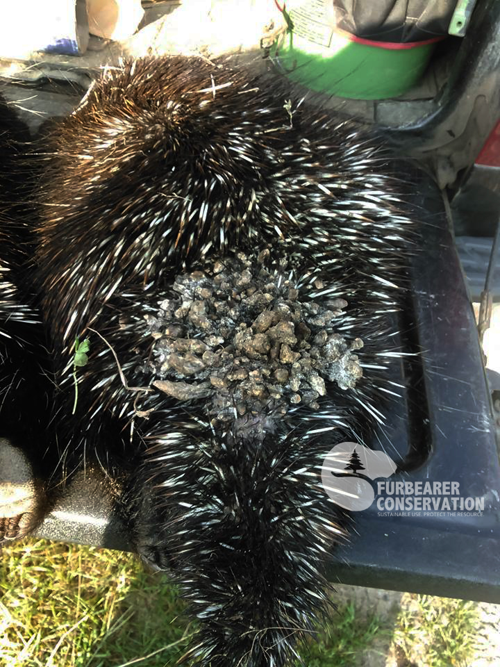
Image furnished by Jeff Traynor (Furbearer Conservation™)
From the Northeast Wildlife Disease Coop:
"Of 44 wild or captive porcupine biopsy or necropsy submissions to the NHVDL from 2010-2017, 28 had significant dermatologic lesions; 12 of these were positive by culture or histopathology for dermatophytes (ring worm). Culture of several recent cases at the NHVDL showed the predominant fungal isolate to be consistent with Trichophyton sp. Molecular analysis of culture isolates at the Department of Population Medicine and Diagnostic Sciences at Cornell University College of Veterinary Medicine identified the predominant dermatophyte to be Arthroderma benhamiae, which is the teleomorph of Trichophyton mentagrophytes (T. interdigitale). This dermatophyte is adapted to an animal hosts (zoophilic), specifically rodents (sylvatic). The lesions are unique to ringworm amongst mammals, as there is a diffuse distribution, mild inflammatory response, dramatic hyperkeratosis, and debilitating to fatal clinical disease in porcupines infected with these fungi.
The primary clinical differential was mange (Notoedres douglasi), which is another common disease in this species. No mites were seen clinically or at necropsy. The degree of hyperkeratosis in this case is comparable to what we have seen in the 5 cases of mange in our collection, but the paws and face tend to be more dramatically affected in cases of mange than dermatophytosis. None of the 12 porcupines with dermatophytosis in our series had concurrent mange. All of the affected animals were found in New England. None of them appeared to be able to fight off the infection. There is currently no information about potential population-level effects of this disease."